CD Laboratory for Ophthalmic Image Analysis


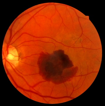
Pathologies of the eye such as age-related macular degeneration, diabetic macular oedema and retinal vascular occlusion are the result of the most common diseases of civilisation and the most important diagnoses in modern Medicine.
As widespread causes of severe and irreversible loss of vision, including blindness, they play a significant role in the care of the population. In line with the change in demographic age distribution, the prevalence of these chronic diseases is increasing massively.
Current pharmacological achievements have made these serious eye diseases extremely effective to treat: various anti-VEGF drugs represent a potent but very resource-intensive treatment option that involves complex interventions (surgical administration of medication directly into the eye) and highly developed, expensive drugs. New developments in diagnostic imaging, in particular non-invasive optical coherence tomography (OCT), allow early detection and targeted monitoring of progression during treatment. Either monthly injections are required or continuous monitoring of the findings using OCT is recommended, with the aim of treating as often as necessary but as little as possible so that the effort, costs and side effects of the therapy can be kept under control. This is because it is currently not possible to determine the optimal treatment time for individual patients. There is a lack of reliable parameters and disease models to identify the individually appropriate treatment period. This laboratory is looking for such parameters and models.
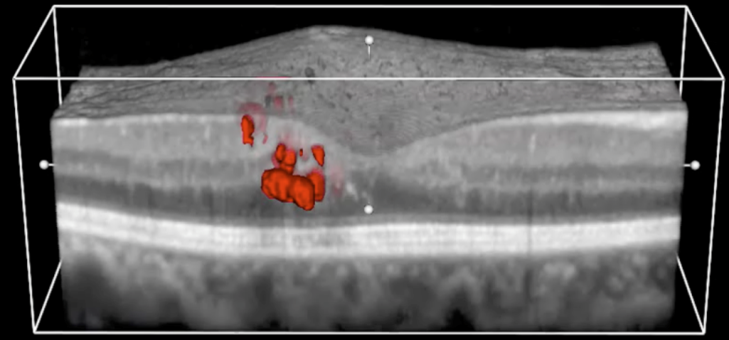
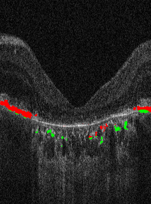
The new generation of OCT (SD-OCT) provides high-resolution and three-dimensional images of the retina. The resolution is so high that individual retinal layers and neurosensory microstructures can be qualitatively assessed; the smallest structures down to cellular changes can be visualised. However, and this is where the need for research lies, this large amount of image data cannot currently be utilised in a meaningful way. On the one hand, it is not known which parameters are actually suitable for predicting the course of the disease, and on the other hand, the large amounts of data need to be processed efficiently.
The aim of the CD Laboratory is to develop new computer-aided methods for analysing images. This should enable standardised and quantitative processing of all information available in OCT so that treatment can be used more efficiently both medically and economically.
An interdisciplinary team works together to develop computer-controlled algorithms for analysing the imaging procedures: Experts in retinal diseases, computer science, medical physics and biomedical optics.
The new methods will not only adapt treatment individually (personalised Medicine), but will also contribute to the identification of comprehensive disease models (population analysis): complex statistical disease models will be developed on the basis of the analysed image data, allowing risk patients to be identified earlier and enabling accurate prognoses to be made.
At the end of the research work, there will be solid standards for one of the most common and most expensive treatment methods in modern Medicine: even after the first examination, it will be possible to recognise how aggressive the disease will be, how many checks and treatments will be necessary, and what treatment outcome can be expected.

Christian Doppler Forschungsgesellschaft
Boltzmanngasse 20/1/3 | 1090 Wien | Tel: +43 1 5042205 | Fax: +43 1 5042205-20 | office@cdg.ac.at

