JR Centre for Materials Engineering in Soft Tissue Regeneration


Tissue engineering plays a central role in regenerative medicine, as it offers solutions for organ failure, tissue damage and degenerative diseases. It also has the potential for personalized therapies. However, there is currently a lack of test and development systems for transferring in-vitro models to living organisms. This JR Centre is researching three-dimensional (3D) bioprinting techniques for use in the regeneration of muscle tissue.
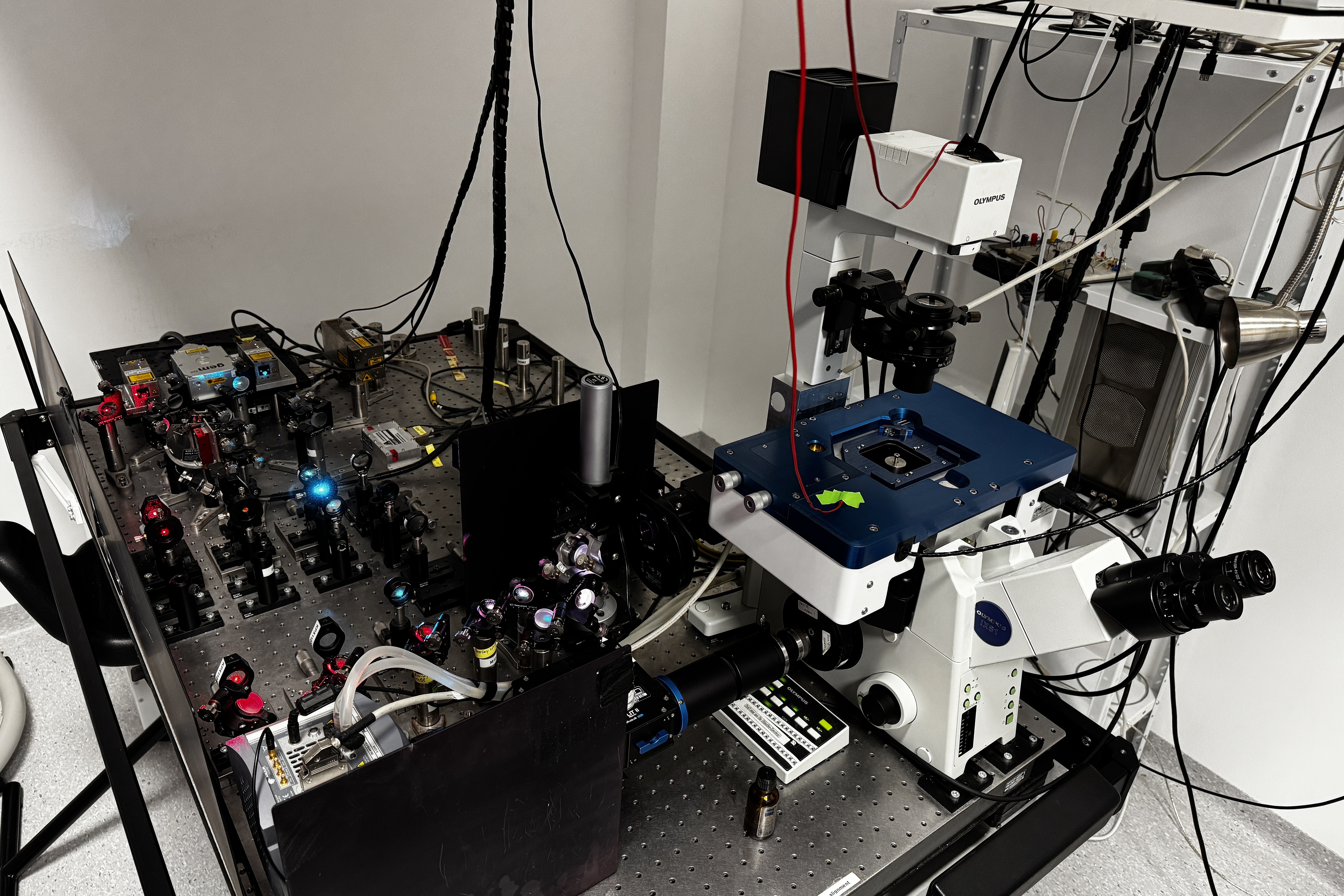
The main objective of the JR Centre is therefore to research how 3D cell scaffolds can be developed from so-called bioinks. These are species-specific (here human and murine), possibly modified proteins of the extracellular matrix (ECM) of muscle tissue. Multiphoton lithography and direct light processing are used as 3D bioprinting techniques. The components of the bioink are polymerized using light. The properties of the ECM proteins are chemically optimized in order to minimize protein damage caused by light absorption and thermal cross-linking. Such printed scaffolds for biological cells are intended to replicate the structure of smooth and skeletal muscle tissue, improve experimental and translational models, enable precise control over geometry and structure and promote personalized tissue replacement.
In order to recreate synthetic muscle tissue, the 3D cell scaffolds must be colonized with muscle cells in the next step. A murine in-vitro cell system for the differentiation of skeletal muscle-derived cells (SMDCs) into functional muscle fibres or transdifferentiated induced smooth muscle cells (iSMCs) will be developed for this purpose. Standardized protocols for mechanical and electrical stimulation will be developed to promote the maturation of 3D muscle models and facilitate the analysis of biochemical and mechanical signals to improve muscle function. In parallel, biomarker tools will be developed to assess the functionality and quality of these models, focusing on muscle gene analysis, ECM proteins and secreted factors such as extracellular vesicles (EV). Furthermore, a pipeline for the quantitative analysis of EV from iSMCs will be developed, especially when enriched from human mesenchymal stem/stromal cells.
In the course of the project, the 3D cell scaffolds will be adapted for pre-clinically relevant muscle defect models in mice, the cell systems will be modified and the composition of the protein-based bioink materials will be further optimized. These muscle defect models are used to evaluate the regenerative properties of the scaffolds colonized with muscle cells. The functionality of the 3D cell scaffolds will be further improved by mechano- and biochemical stimulation (e.g. EV addition) to increase in-vivo tissue mimicry and ensure implantation compatibility. The developed tool set will be versatile for the regeneration of numerous other soft tissues.

Links
Christian Doppler Forschungsgesellschaft
Boltzmanngasse 20/1/3 | 1090 Wien | Tel: +43 1 5042205 | Fax: +43 1 5042205-20 | office@cdg.ac.at

