CD Laboratory for Clinical Molecular MR Imaging
Head of research unit
Commercial Partner
Duration
Thematic Cluster


This CD Laboratory develops high-resolution, quantitative imaging techniques for early, specific diagnosis and prompt evaluation of therapy response. Both significantly improve the prognosis of patients.
Many diseases can only be diagnosed when morphological changes come to light. These are typically visualised using ultrasound, computer tomography or magnetic resonance (MR) tomography. However, in order to improve the prognosis of patients with the disease, diagnostics are required that can detect molecular changes even before morphological manifestations occur. Due to the diversification and personalisation of available forms of therapy, there is also a need for therapy progress monitoring that can detect an individual response or non-response at an early stage.
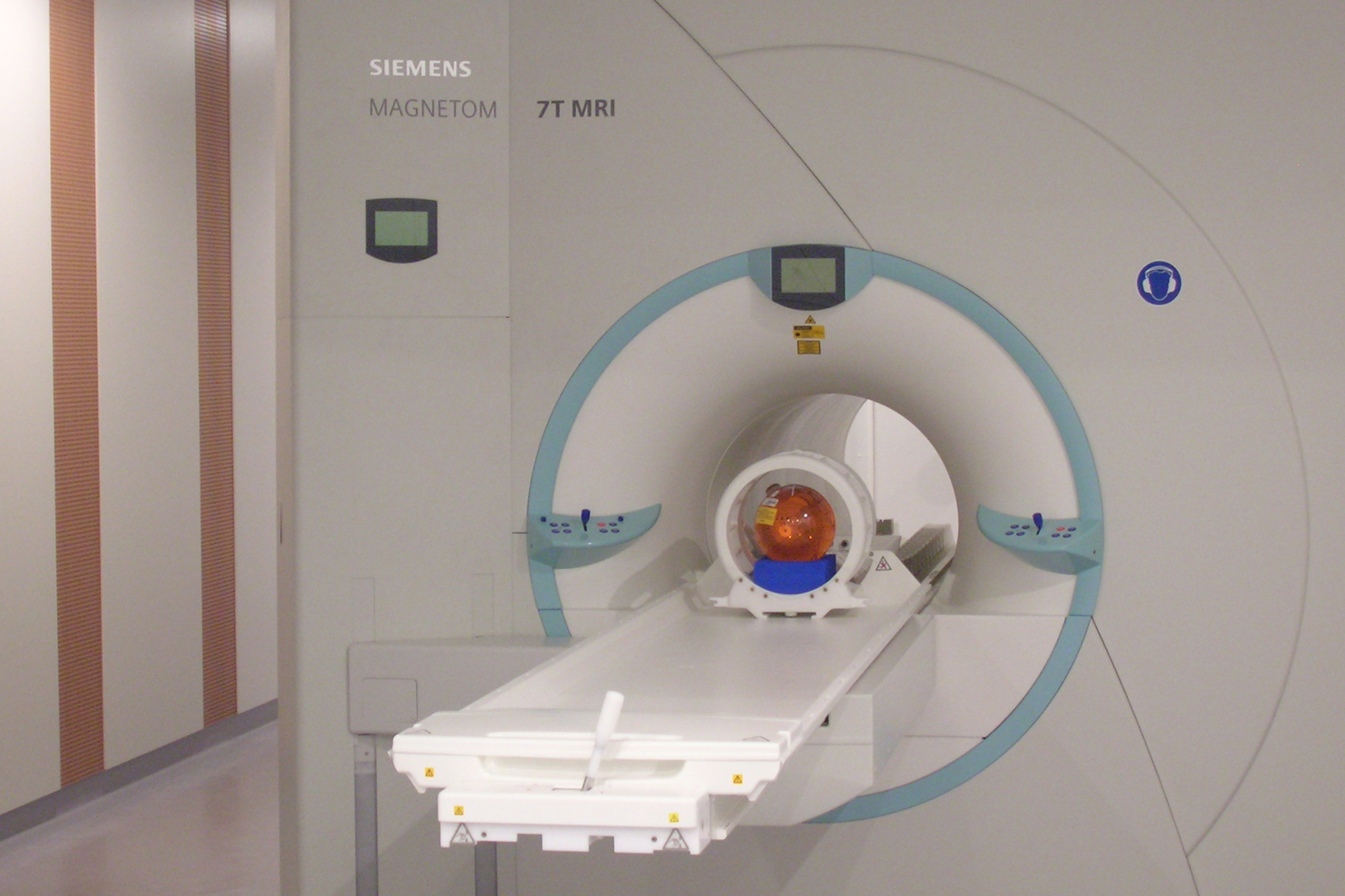
This CD Laboratory for Clinical Molecular MR Imaging (MOLIMA) develops methods of MR spectroscopy (MRS) in the high-field and ultra-high-field range with 3 and 7 Tesla flux density, respectively, which can provide the required resolution and sensitivity in the molecular range. Such powerful magnets have a better signal to background noise ratio resulting in higher spatial resolution, higher sensitivity and higher spectral resolution. Atomic markers that can be specifically observed in this way include protons as well as other nuclei that are much rarer in the human body, such as phosphorus-31 and sodium-23. Molecular groups can be quantified using chemical exchange saturation transfer (CEST) imaging.
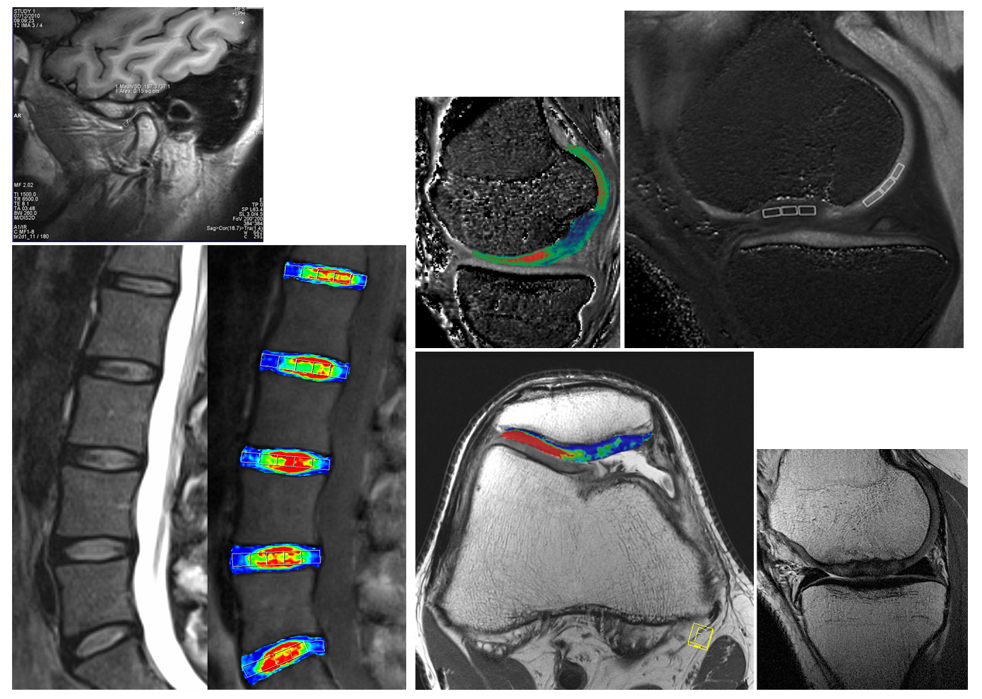
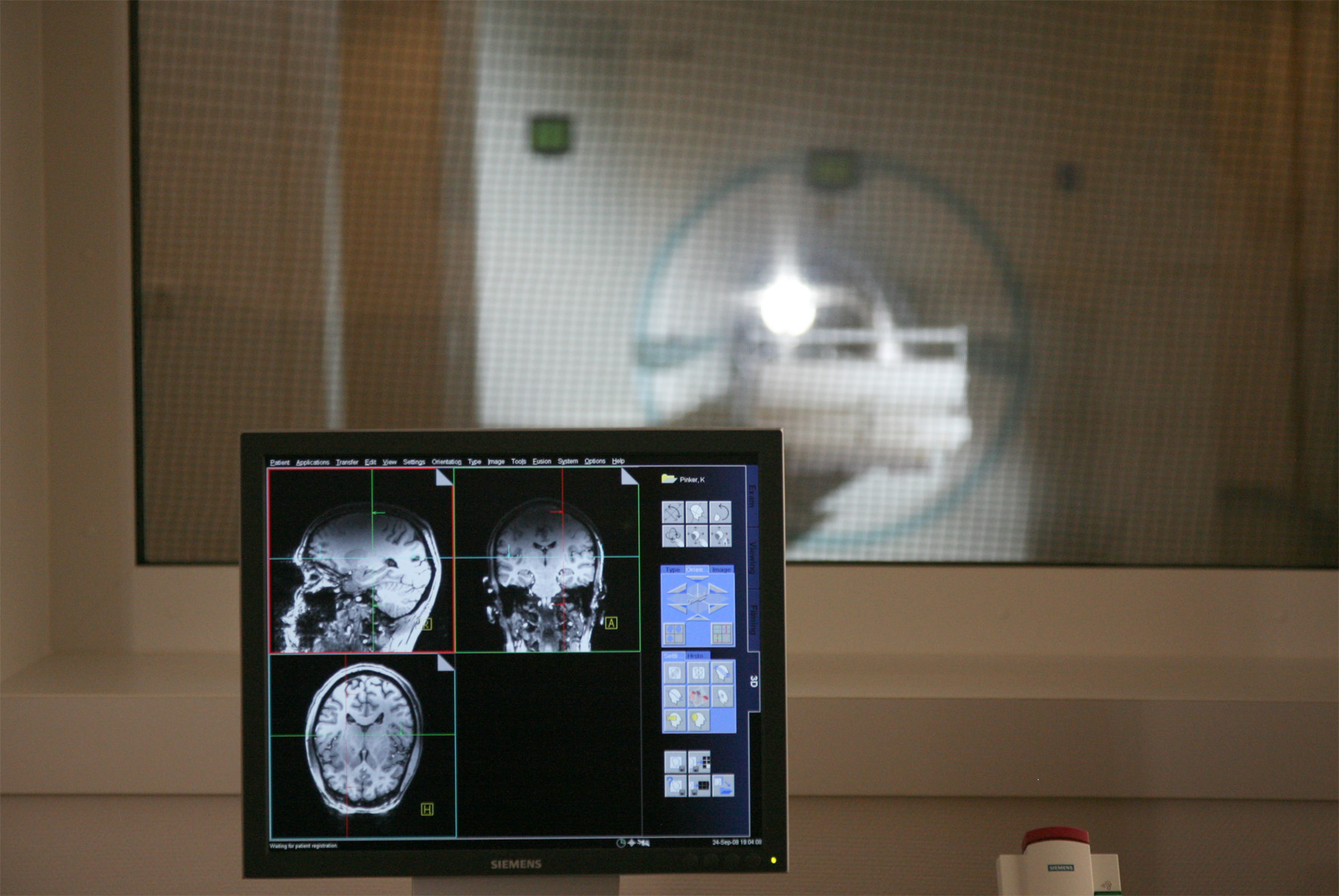
Proton-based MRS is used to analyse metabolic processes in the entire brain. Using phosphorus-31 MRS, signals from several muscle groups can be recorded simultaneously and their performance analysed individually. Performance is measured as the capacity for mitochondrial synthesis of ATP from inorganic phosphate (Pi) and can be carried out non-invasively even on actively moving muscles. Sodium-23 MRS is used for imaging the kidneys, the musculo-skeletal area (cartilage, ligaments, intervertebral discs) and breast cancer and selectively measures the intracellular sodium concentration as well as the total sodium concentration. CEST imaging is used in particular to characterise larger molecules. Changes in cartilage are determined via its glycosaminoglycan content, changes in muscle tissue via the creatine content and breast tumours via the choline content.
The aim of this CD Laboratory is to make the methods described suitable for clinical application in various regions of the body and for various diseases and to establish the CD Laboratory as a worldwide reference centre for the clinical application of MRS on 7 Tesla devices.


Christian Doppler Forschungsgesellschaft
Boltzmanngasse 20/1/3 | 1090 Wien | Tel: +43 1 5042205 | Fax: +43 1 5042205-20 | office@cdg.ac.at

